When Should You Get a Chest X-Ray for Heart Disease?
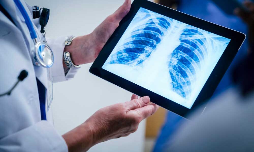
A chest X-ray is one of the tools your doctor may use to diagnose heart disease. This procedure uses a small amount of radiation to create an image of your heart, lungs, and blood vessels.
Why Your Doctor Recommends a Chest Imaging Test
Doctors order chest X-rays to:
- Examine your chest bones, heart, and lungs.
- Verify the placement of heart devices like pacemakers or defibrillators.
- Check the position of any catheters or chest tubes.
Preparing for a Chest X-Ray
You don’t need special preparation for a chest X-ray. However, let the technician know if you could be pregnant. The procedure usually lasts 10-15 minutes. You’ll need to remove clothing and jewelry from the waist up and wear a hospital gown. Standing still while holding your breath is important during the imaging.
How the Process Works
You’ll press your chest against the cassette that holds the film, while the machine sends X-rays through your body. The rays pass through the chest, creating a picture on the film. Dense areas, like bones, appear lighter, while less dense areas, such as the lungs, appear darker. You will then repeat the process with your side against the cassette.
What the X-Ray Reveals
A chest X-ray helps your doctor detect:
- Fluid in or around your lungs
- An enlarged heart
- Blood vessel issues
- Congenital heart disease
- Calcium buildup in the heart or blood vessels
By using this simple and painless test, your doctor can gather valuable information to determine the next steps in diagnosing and treating heart disease.
At Carepoi, we aim to provide accessible healthcare solutions, including advanced diagnostic tools, to ensure quality patient care.


